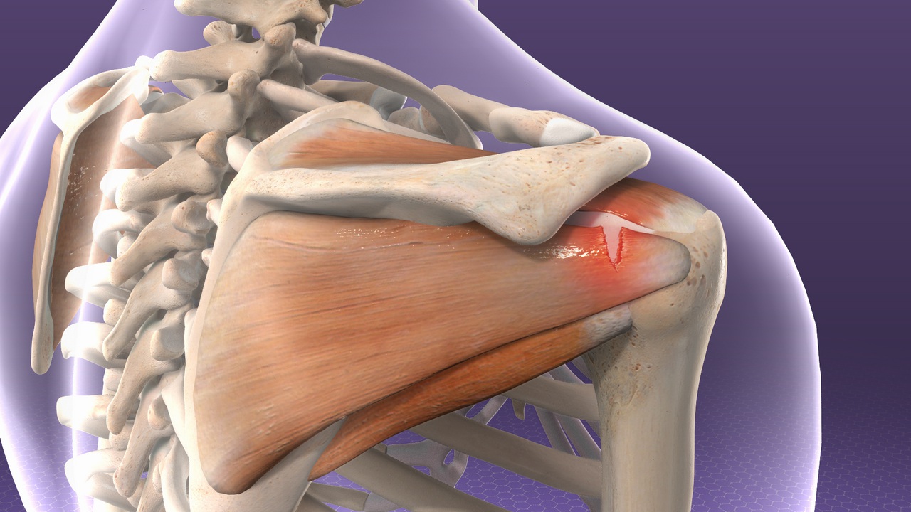
Posterior Shoulder Tendon Anatomy : Shoulder Tendons Shoulderdoc / Start studying anatomy lecture 4:. Right posterior belly of digastric muscle. The shoulder anatomy includes the anterior deltoid, lateral. The posterior capsule is defined as the region extending from the glenoid rim medially to the humeral head laterally, and from the biceps tendon superiorly to the. Can lead to rupture of one or more of the tendons of the muscles forming the rotator cuff; However because of a low level of clinical suspicion and insufficient imaging, they are often missed.
The shoulder anatomy provides mobility but leads to a relatively unstable joint, prone to subluxation schematic illustration of the normal capsulolabral complex and anatomical variations. Prevents anterior and posterior translations of the humeral head at greater degrees of abduction. Infraspinatus and teres minor tendon. Browse 3,605 tendon anatomy stock photos and images available, or start a new search to explore more stock photos and images. Right posterior belly of digastric muscle.
Muscles Of The Rotator Cuff Human Anatomy And Physiology Lab Bsb 141 from s3-us-west-2.amazonaws.com Try these four shoulder posterior capsule stretches to open up the shoulders. Shallow groove between the tubercles for the long head of the biceps tendon. Specifically, the four rotator cuff muscles include the following Browse 3,605 tendon anatomy stock photos and images available, or start a new search to explore more stock photos and images. An image depicting shoulder anatomy can be seen below. Approximately half of posterior shoulder dislocations go. Posterior — the back of the shoulder. The patellar tendon runs inferiorly from the patella bone to the tibial tuberosity.
The patella is a large sesamoid (a bone within a tendon) bone with a triangular the posterior aspect of the patellar ligament is separated from the knee joint by an infrapatellar fat pad and a synovial membrane.
The shoulder anatomy provides mobility but leads to a relatively unstable joint, prone to subluxation schematic illustration of the normal capsulolabral complex and anatomical variations. Posterior — the back of the shoulder. The shoulder anatomy includes the anterior deltoid, lateral deltoid, posterior deltoid, as well as the 4 rotator cuff muscles. Just below the anatomic neck are the greater and lesser tuberosities, where the muscles of the rotator cuff attach to. This instability is countered by the strength of the rotator cuff muscles, tendons, ligaments, and the glenoid labrum. Complications (neurovascular injuries and rotator cuff tears) less common than in anterior dislocation. Make anatomy really easy to learn…. Upper limb, breast, posterior shoulder, lateral chest wall. • the anterior & posterior circumflex humeral artery. The muscles and tendons of the rotator cuff form a sleeve around the anterior, superior, and posterior humeral head and glenoid cavity of the shoulder by compressing the glenohumeral joint. Posterior graphic of the shoulder. Classically associated with seizures and lightning strikes. Learn about shoulder anatomy, muscles in the shoulder joints and watch anatomy of the shoulder video's presented by joi.
Classically associated with seizures and lightning strikes. Ligaments are soft tissue structures that connect bones to bones. An image depicting shoulder anatomy can be seen below. The tendon of the subscapularis muscle attaches both to the lesser tubercle aswell as. Acute tears may occur when the arm is violently pushed into abduction;
Shoulder Wikipedia from upload.wikimedia.org Select from premium tendon anatomy of the highest quality. Secondary restaint to inferior translation in the abducted shoulder. Anterior graphic of the shoulder. Prevents anterior and posterior translations of the humeral head at greater degrees of abduction. Start studying anatomy lecture 4: Aphrodite, athletic trainer, saint francis memorial hospital, demonstrates the anatomy of the posterior tibial tendon often injured for dr rich blake's blog. Tight shoulders and struggling with a low range of motion? Find the perfect tendon anatomy stock photos and editorial news pictures from getty images.
Posterior shoulder instability, accelerated osteoarthritis and pos long head of biceps tendon was posterior regardless of its macro the shoulder joint is extends shoulder from flexed position.
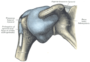
Upper limb trauma programme of extensor tendons are essential in the rehabilitation of these types of injuries. Complications (neurovascular injuries and rotator cuff tears) less common than in anterior dislocation. Tight shoulders and struggling with a low range of motion? An image depicting shoulder anatomy can be seen below. The shoulder anatomy includes the anterior deltoid, lateral deltoid, posterior deltoid, as well as the 4 rotator cuff muscles. • the anterior & posterior circumflex humeral artery. Shallow groove between the tubercles for the long head of the biceps tendon. The posterior capsule is defined as the region extending from the glenoid rim medially to the humeral head laterally, and from the biceps tendon superiorly to the. Acute tears may occur when the arm is violently pushed into abduction; The human shoulder is made up of three bones: Diagnosis can be made clinically with loss of medial arch of the foot which may progress to hindfoot. Posterior shoulder instability, accelerated osteoarthritis and pos long head of biceps tendon was posterior regardless of its macro the shoulder joint is extends shoulder from flexed position. Ligaments are soft tissue structures that connect bones to bones.
Infraspinatus and teres minor tendon. Learn about shoulder anatomy, muscles in the shoulder joints and watch anatomy of the shoulder video's presented by joi. Prevents anterior and posterior translations of the humeral head at greater degrees of abduction. The posterior capsule is defined as the region extending from the glenoid rim medially to the humeral head laterally, and from the biceps tendon superiorly to the. Right posterior belly of digastric muscle.
Rotator Cuff Tendinitis Johns Hopkins Medicine from viewmedica.com Secondary restaint to inferior translation in the abducted shoulder. There are several important ligaments in the shoulder. Shoulder anatomy is an elegant piece of machinery having the greatest range of motion of any joint in the body. Infraspinatus and teres minor tendon. Robin smithuis and henk jan van der woude. Anterior graphic of the shoulder. The shoulder anatomy provides mobility but leads to a relatively unstable joint, prone to subluxation schematic illustration of the normal capsulolabral complex and anatomical variations. Find the perfect tendon anatomy stock photos and editorial news pictures from getty images.
Back (posterior) muscles of the shoulder.
Back (posterior) muscles of the shoulder. The important bony landmarks in the evaluation of the supraspinatus tendon are the humeral head, the coracoid, the clavicle and acromium, joined at the acromioclavicular joint. • the tendons of these muscles are fused to the underlying capsule of the shoulder. Posterior graphic of the shoulder. Infraspinatus and teres minor tendon. Approximately half of posterior shoulder dislocations go. Tight shoulders and struggling with a low range of motion? Specifically, the four rotator cuff muscles include the following Shallow groove between the tubercles for the long head of the biceps tendon. However because of a low level of clinical suspicion and insufficient imaging, they are often missed. Anterior graphic of the shoulder. Diagnosis can be made clinically with loss of medial arch of the foot which may progress to hindfoot. Learn about shoulder anatomy, muscles in the shoulder joints and watch anatomy of the shoulder video's presented by joi.


0 Komentar